
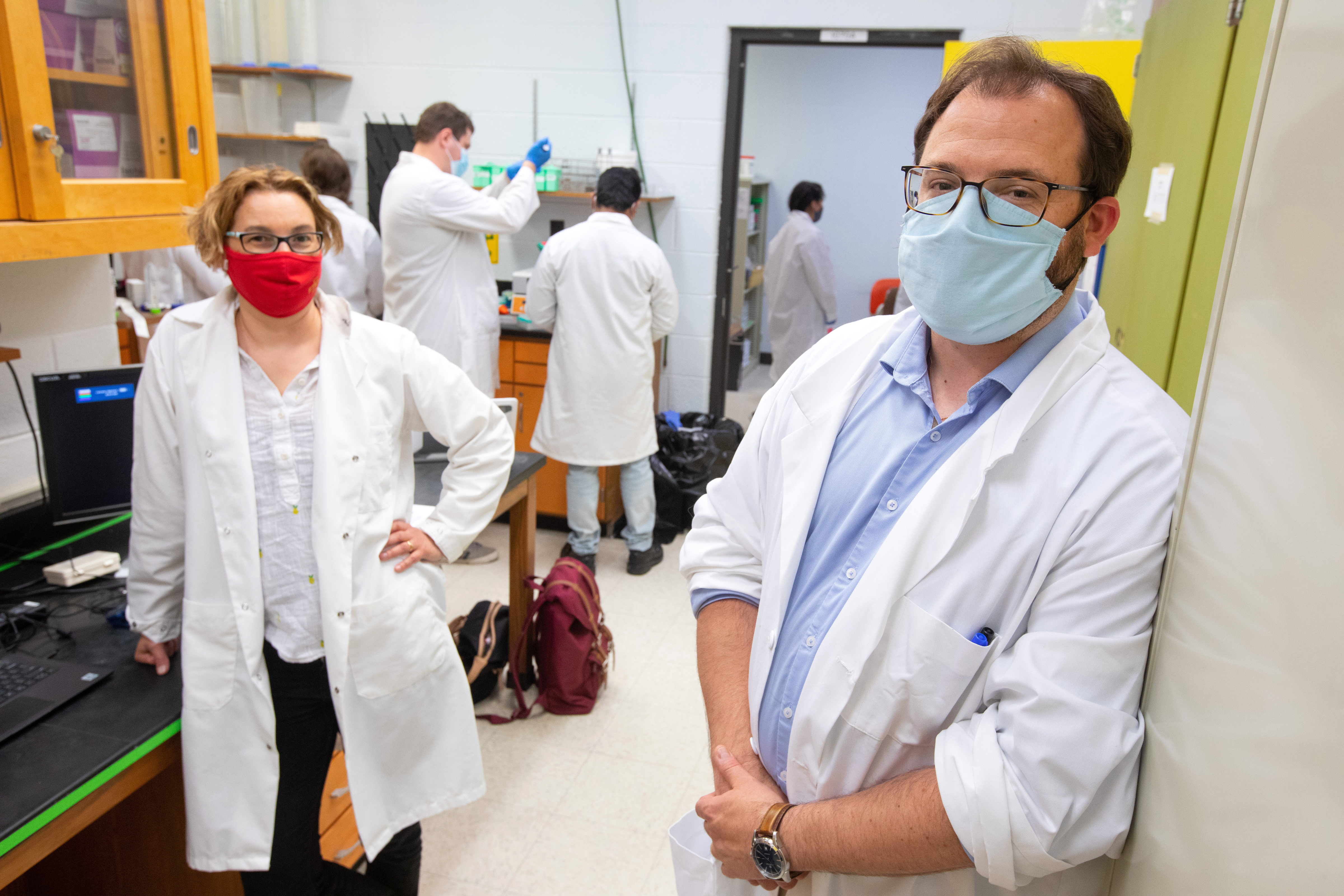
Karin Allesnpach, left, and Jon Mochel, right, and members of their lab are perfecting a means of culturing organoids from patient urine samples on which to test bladder cancer treatments. They say their method has a number of advantages that could lead to better outcomes for patients. Photo by Christopher Gannon. Larger image.
AMES, Iowa – Bladder cancer presents a number of challenges when choosing a course of treatment, but researchers at Iowa State University hope their innovative research might help doctors and patients arrive at an optimal treatment plan faster.
Doctors and patients must weigh numerous variables when choosing the right course of treatment following a bladder cancer diagnosis, and it can be difficult to predict how patients will respond. ISU scientists are perfecting an innovative new technique that grows organoids from patients’ urine samples on which various treatments can be tested. This approach could allow doctors to tailor therapies to patients without the need for invasive surgeries, which often take significant lengths of time to schedule.
The research group includes Jonathan Mochel, an associate professor of biomedical sciences; and Karin Allenspach, a professor of veterinary clinical sciences. Their team has begun a clinical trial of their method in conjunction with physicians at the Mayo Clinic. And earlier this year, they published an article in the peer-reviewed scientific journal Cancers detailing their methods.
They’ve also partnered with Coralville-based biomedical company NanoMedtrix to test their method in dogs, an effort that received support from the National Institutes of Health this year.
Organoids derived from urine samples
The researchers’ technique begins with a urine sample from a patient suspected of having bladder cancer. The researchers use a centrifuge to isolate tumor cells from the urine. Next, they combine the tumor cells with Matrigel, a gelatinous protein mixture in which organoids grow and behave similarly to the patient’s tumor. Doctors then use these tumor-derived organoids as models for patient tumors. Researchers can test treatments, such as various forms of chemotherapy, and gauge the organoid’s response. This gives doctors critical clues as to how a patient’s tumor may respond to treatment in the real world, data that medical professionals can use to devise an individualized treatment plan in patients.
“The idea of precision medicine, or personalized medicine, is central here,” said Allenspach. “It’s hard to predict which people will respond to what treatment. Culturing these organoids helps us to predict which therapeutic the patients will respond to.”
Growing organoids in this manner usually requires cells taken directly from the tumor via surgery. Patients sometimes must wait weeks or months to schedule a biopsy with a surgeon but deriving the organoids from urine samples is a novel and non-invasive approach that could be carried out much faster. That means doctors can determine a course of treatment earlier, leading to better patient outcomes, Allenspach said.
Clinical trials in dogs and humans
The NIH grant, awarded through the National Cancer Institute, will fund clinical trials in dogs with bladder cancer. In addition, and through their partnership with NanoMedtrix, a company that specializes in nanomaterials, the researchers aim to enroll 18 dogs over two years to receive experimental therapies for bladder cancer. NanoMedtrix has developed particles that improve how medications bind to cancer cells in the bladder so less of the medicine is flushed out in the patient’s urine. The company concluded an earlier study of their particles in mice and are teaming up with Iowa State to see how the approach works in dogs. In doing so, Mochel and Allenspach will test their method of developing organoids from urine samples to guide the experimental treatments.
Using dogs as a model offers insights that could greatly speed up that development of new treatments because many of the diseases they suffer, such as bladder cancer, are virtually identical to humans, making them better models than rodents, Mochel said.
Dog owners who think their pets might be good candidates for the trials can apply trough the college’s clinical trial service website.
The researchers started clinical trials in humans in May in collaboration with doctors at the Mayo Clinic in Rochester, Minnesota. The trials will involve up to 200 patients with bladder cancer. The ISU researchers will receive urine samples from the patients and use organoids derived from the samples to predict what kind of treatments will be most effective. Unlike in the clinical trials in dogs, however, the researchers’ findings will not be used to make treatment decisions for the human patients until the method has been validated against clinical outcome in people.
The bladder cancer research builds on work Mochel and Allenspach have conducted to test new drugs on organoids derived from canine stem cells. Just as in their bladder-cancer models, the researchers have devised a way to grow clusters of canine cells that mimic dog intestines. The researchers use this model to test how well new drugs are absorbed by canine intestinal tissues, an important indicator of how effective a drug will be as a treatment. That’s key because drug companies can spend more than a decade and millions of dollars in the development of new treatments, only to find the new drugs aren’t effective in humans.
Testing new drugs on 3D organoids can provide earlier indications of whether a new experimental drug is sufficiently efficacious and safe to continue testing and development, Mochel said.