New dynamic knee anatomy model helps athletic training students identify meniscus injuries
February 16, 2026
AMES, Iowa – A distinct popping sound or sudden catch in movement.
An athlete’s expression of sharp, intense pain.
In the field of athletic training, understanding the human body isn’t optional – it’s the foundation of every evaluation, diagnosis and treatment decision that shapes a patient’s recovery, which begins in the moments following an injury.
Some structures in the body, however, can be difficult to fully understand using textbooks and static models alone, potentially making certain injuries a bit more complicated, too. One notoriously tricky example is the knee meniscus, said Megan Brady, clinical assistant professor of kinesiology and health at Iowa State.
“The meniscus can have a variety of tear patterns, and the subtle differences between these injuries can make it hard for students to visualize what’s really happening inside the knee,” said Brady, who is also a core athletic training faculty member at ISU.
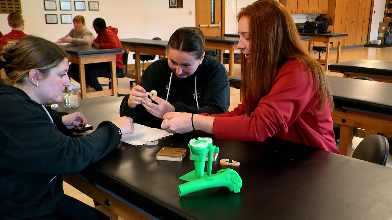
It was this challenge that led Brady to design and fabricate a new dynamic knee anatomy model that captures the motion and inner workings of the knee – and by extension, improves student learning and understanding of the meniscus.
“Anatomy has historically been taught in three dimensions using dissection, surface anatomy and static models, and in two dimensions using textbooks, drawings or digital images,” Brady said. “I was excited to see if we could create something different and improve visualization in a real and meaningful way.”

In collaboration with the ISU Student Innovation Center and Dan Neubauer, a former associate teaching professor of industrial design at Iowa State and current associate teaching professor at the University of Wisconsin-Stout, Brady worked to design and fabricate a 3D-printed knee anatomy model of bones and meniscus using plastisol.
The plastisol, a liquid substance that can be converted into solid plastic, was heated and poured into a 3D-printed meniscus mold before being placed on a 3D-printed tibia to solidify.
And the best part?
“The plastisol meniscus can be cut, reheated and remolded over and over,” Brady said, “which gives us the ability for varied and ongoing use in our classrooms and labs.”
Brady said the goal of this project was to create a dynamic knee anatomy model that would allow educators to demonstrate various types of meniscus tears and help their students better visualize and understand how those various tears may impact each individual patient – including why some meniscus tears require surgery while others do not.
The knee itself is made up of four bones: the femur (or thighbone) is the bone that connects the hip to the knee; the tibia (or shinbone) is the bone connects the knee to the ankle; the patella (or kneecap) is the small bone in front of the knee and rides on the knee joint as the knee bends; and the fibula (or calf bone) is a shorter and thinner bone that runs parallel to the tibia on its outside and acts as a crucial support structure.
“The knee has two C-shaped pieces of cartilage – which are called meniscus – that act like a cushion between the tibia and femur,” Brady said. “And a torn meniscus is one of the most common knee injuries because any activity that causes you to forcefully twist or rotate your knee, especially when you’re putting your full weight on it, can lead to a tear.”
Types of meniscus tears include radial, horizontal, flap, complex and bucket-handle, and while they each can compromise the knee, Brady said, the part of the meniscus these tears affect – as well as the patterns they exhibit and their complexity – differ.
“Meniscus tears can be difficult to identify because symptoms like pain and swelling often come and go, and some tears can even be asymptomatic,” Brady said. “So, with the dynamic knee anatomy model, I felt that if I could find something that moves and if I could demonstrate, ‘here’s the mechanism and here’s the result of the mechanism,’ it would help give students a better idea of what’s going on in the knee and realistic steps for treatment.”

Mary Meier, clinical professor of kinesiology and health at Iowa State, said she was excited when Brady came to her with the idea to create the new dynamic knee anatomy model.
“As a health care professional and as an athletic training clinician, we’re always trying to find the best ways to take care of our patients and explain injuries to them, and when we teach our students, we want to be able to show those methods to them and really demonstrate what we’re talking about in relation to a particular injury,” said Meier, who is also director of ISU’s athletic training program.
Morgan Severseike, a second-year graduate student in Iowa State’s 3+2 athletic training program, said the new dynamic knee anatomy model has been a helpful learning aid, especially when it comes to full visualization of the knee.
“A textbook can only teach you so much and you can’t really take off someone’s leg to look at a knee injury, so being able to see that model is really helpful,” Severseike said.
Khalil Jeter, another second-year graduate student in Iowa State’s 3+2 athletic training program, also credited the new model with providing a quicker understanding of meniscus tears.

“Once you see (the model) in person and you can see how the meniscus is moving, you’re understanding how these tears can happen and visualizing what they look like in real time,” Jeter said.
Brady said the dynamic knee anatomy model is now being used in lower-body evaluation courses to help provide ISU athletic training students with a solid understanding of several types of meniscus tears, and she plans to continue refining the design and additional usage possibilities.
“Innovative instructional tools can take learning to a new level and help prepare our students to excel in the workforce,” Brady said.
Iowa State’s athletic training program is a professional master’s degree program, and students completing their third year of Iowa State’s 3+2 athletic training program, as well as students who have completed a B.A. or B.S. degree from an accredited institution, are eligible to apply. Students in the program interact with team physicians, chiropractors, orthopedic surgeons, physical therapists and other health care providers as part of the program’s comprehensive, hands-on clinical experiences.